Delve Deeper: Understanding the Neuroscience behind Disaster Recovery (Part 2 of 3)
Nicole Gunawansa | July 7th, 2015
This interview was held on April 21st, 2015 in the IDAC "Smart Aging" Building
Part 2- Disaster Resilience: Neurocognitive Perspective
Question 4: Please explain the relationship between brain structure/chemistry and resilience after 3/11. What research is currently being conducted by your lab in this area?
Dr. Sekiguchi: We have published papers related to this topic using data from our previous lab (Dr. Kawashima’s lab) ("Resilience after 3/11: structural brain changes 1 year after the Japanese earthquake" and "Brain structural changes as vulnerability factors and acquired signs of post-earthquake stress"). Prior to the earthquake we used brain MRI database in IDAC to store information from healthy Tohoku University student participants. After the earthquake, we re-recruited these students (now survivors of the disaster) to assess risk factors with a neural basis for PTSD symptoms soon after the disaster. This study evaluated longitudinal changes, and we were able to detect subclinical vulnerability factors and occurrence of PTSD symptoms. We followed up with our participants one year after the disaster, performing brain imaging and questionnaire based assessment of PTSD symptoms. This follow up data demonstrated that PTSD symptoms recovered significantly. Small psychological changes which occurred immediately before or after the disaster resulted in shrinking prefrontal cortex gray matter. One year later, this reduced brain volume recovered in correlation with increased levels of self-esteem, indicating resilience in specific populations. The research for the study described is finished because student participants have graduated. In the future, we hope to replicate the previous study and asses the role of self-esteem in brain recovery from disaster distress as a part of a large scale ToMMo cohort study.
Question 5: How does your department encourage public participation for the MRI studies? Are there specific qualification/criteria for participation?
Dr. Sekiguchi: Since our current MRI study is a part of ToMMo’s cohort analysis, we recruit healthy subjects from the Community-Based Cohort Study and the Birth and Three-Generation Cohort Study. We take those who are willing to join, and send documentation to those people already recruited in the cohort. Potential participants must be over the age of twenty. Participants cannot contain metal or foreign body in/on their person (unless approved by a physician). This criterion is very important because MRI radio waves cause electric power and current, which can result in the heating of metal/foreign body during MRI scanning, potentially harming the participant. We have the participants’ best interest in mind, and will only take participants with metal (i.e. from a surgical procedure) whose physicians provide permission to join in the study. Additionally, we do not allow pregnant women or those with tattoos in the study because of potential harm to the fetus and the small amounts of metal (iron) in tattoos respectively.
Dr. Taki: We are collecting research data from relatively healthy subjects (having no serious or terminal aliments), and thus we have very strict exclusion criteria for this study.
Question 6: What is the role of MRI in post natural disaster evaluations? Do you think the use of MRI in disaster recovery will continue to grow in the future?
Dr. Taki: Clinically, the prevalence of diseases such as diabetes, hypercholesterolemia, and hypertension has increased due to stress and changes in nutrition following the disaster. These disorders increase risk of cerebral infarction (type of ischemic stroke resulting from blockage in the blood vessels supplying blood to the brain), but MRI helps to combat this risk with the early diagnosis and prevention. Aside from the clinical aspect, we hope do expand our research (in the future) to include different types of trauma in order to document differences in affected brain areas, potentially resulting in various structural and functional changes. Utilization of this knowledge can advance both clinical and research disaster medicine, and allow for the creation of more specialized disaster mental health 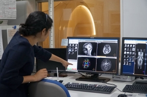 practices.
Dr. Sekiguchi: In current research, MRI is primarily used for macroscopic diagnosis. Although we cannot detect disorder with just visual imaging, use of MRI analysis allows us to see many structural changes as well; thus, MRI proves to be very useful for diagnosis and assessment of recovery. In the future, we can use MRI for clinical and end phenotype diagnosis, detection of risk factors, and promotion of early clinical intervention before the manifestation of these disease. However, it is important to note that MRI imaging for survivors is challenging, and so alternatives for disaster recovery are imperative.